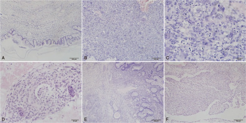
Figure 1.

Microscopic appearance of ovarian tumor and greater omental lesion (hematoxylin-eosin staining). A: A mucinous cystadenoma is the main part of the epithelial elements (×100). B: The sarcomatous mural nodule consists of ovoid mononucleated cells and numerous multinucleated osteoclast-like giant cells (×100). C: Nine mitotic figures are observed in one high-power field in the mural nodule consisting of pleomorphic undifferentiated sarcoma (×400). D: Vascular invasion is observed in the lager mural nodule (×200). E: Benign Brenner tumor is composed of nests of transitional epithelium (×100). F: The greater omental lesion shows nodular histiocytic aggregates.
