Figure 2.
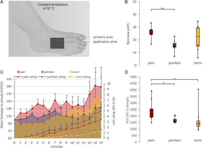
Capsaicin challenge and vasoactive reaction. (A) Setup of the experimental capsaicin application procedure. In the application area, the capsaicin patch was applied and the sensory testing was conducted. In the primary area, the continuous blood perfusion measurement as well as the margin of the axon reflex erythema was determined. (B) The axon reflex flare area (cm2) after 15 minutes of topical capsaicin application is displayed as boxplot (minimum, maximum, median, and first and third quartiles). There were no significant differences between the patient groups and healthy subjects. Capsaicin induced a larger axon reflex flare in the neuropathic pain patients than in the patients without pain. The application area (9 cm2) was not included in the total axon reflex flare size. Interindividual statistical testing for axon reflex flare area was conducted using the Mann–Whitney U test. **<0.01. (C) Displayed is the time course of mean (±SEM) blood perfusion change (arbitrary perfusion units, % change [PU%]) in the primary area and the mean (±SEM) pain rating change (numeric rating scale 0–10) during capsaicin challenge. Interindividual statistical testing for maximum pain ratings (ie, at minute 15) was conducted using the Mann–Whitney U test (statistically not significant). (D) The area under the curve for blood perfusion (AUC [PU%]) is displayed as boxplot. Indicated is a significant difference between the patients with pain as compared to the painless and normal subgroups. Interindividual statistical testing for mean blood perfusion (AUC) between subgroups was conducted using the Mann–Whitney U test. *<0.05.

