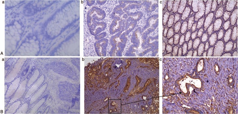
Figure 4.

Immunohistochemical analyses of pERK and c-inhibitor of apoptosis protein 2 (c-IAP2) expression. Higher pERK (A) and c-IAP2 (B) protein expression was observed in colorectal cancer tissues, compared to the adjacent normal mucosa (magnification: a and b, 100×; c, 200× SP). (A-a and 4B-a) Normal tissue. (A-b, B-b) Tumor tissues in Dukes stage A-B. (A-c and B-c) Tumor tissues in Dukes stage C-D.
