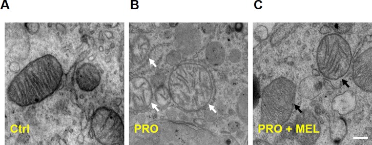
Fig 6. Transmission electron microscopic examination of ultrastructural morphology of mitochondria in microglia.
Figures showed representative electron micrographs of microglia. The left column showed normal ultrastructural features of mitochondria (A), White arrows pointed to mitochondria with “dissolving” features in prorenin-stimulated microglia (B), and the right column showed melatonin cotreatment protected the normal ultrastructural morphology of the mitochondria (C), which revealed that melatonin markedly alleviated mitochondrial swelling, cristae disorientation and breakage compared with the prorenin-treated group. Scale bar = 1 nm.

