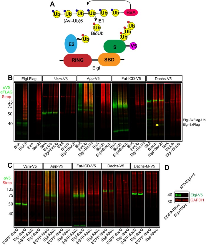
Fig 5. elgi does not ubiquitinate Vam, App, Fat-ICD or Dachs.
(A) Schematic illustrating the ubiquitination assay used. BirA covalently conjugates Biotin to the fused Avi-tagged ubiquitin, which is processed by the E1 to produce biotinylated ubiquitin (BioUb), which is subsequently incorporated by the ubiquitination machinery onto the substrates. This can be detected by fluorescently conjugated streptavidin. (B) Western blot showing results of ubiquitination experiments on proteins expressed in S2 cells. For Elgi autoubiquitination, 3xFLAG tagged Elgi was co-expressed with either BirA or BirA-BioUb and immunoprecipitated using anti-Flag beads. Elgi was detected by anti-FLAG antibody. V5-tagged Full length Vam (Vam-V5), App (App-V5), Fat-ICD (Fat-ICD-V5) or Dachs (Dachs-V5) were co-expressed with either BirA alone or BirA-BioUb with or without Elgi-3xFLAG. V5-tagged proteins were immunoprecipitated from cell lysates, and detected with anti-V5 antibody. In all lanes, biotinylated Ubiquitin was detected with IRDYE680 conjugated streptavidin (Red). The yellow arrow points to a faint band of Elgi-3x-FLAG immunoprecipitated with Dachs:V5; the strong red band just above it is ubiquitinated Elgi-3x-FLAG, based on size and detection with anti-FLAG antibodies. Its appearance presumably reflects the strong association of Elgi with Dachs. (C) Western blot showing results of ubiquitination experiments on proteins expressed in S2 cells treated with dsRNA against either EGFP (control) or elgi. V5-tagged Full length Vam (Vam-V5), App (App-V5), Fat-ICD (Fat-ICD-V5), full length Dachs (Dachs-V5) or middle domain of Dachs (D-M-V5) were co-expressed with BirA-BioUb. V5-tagged proteins were immunoprecipitated from cell lysates and detected with anti-V5 antibody; Biotinylated Ubiquitin was detected with IRDYE680 conjugated streptavidin (Red). (D) Western blot showing efficiency of dsRNA-mediated depletion of elgi in S2 cells. V5-tagged Elgi (Elgi-V5) was expressed using MT-Gal4 with 0.5mM CuSO4 in presence of 10uM MG132 in S2 cells treated with dsRNA against either EGFP (control) or elgi, and whole cell lysate was examined for expression of Elgi-V5 with Rabbit anti-V5 antibody. GAPDH was used as a loading and transfer control.

