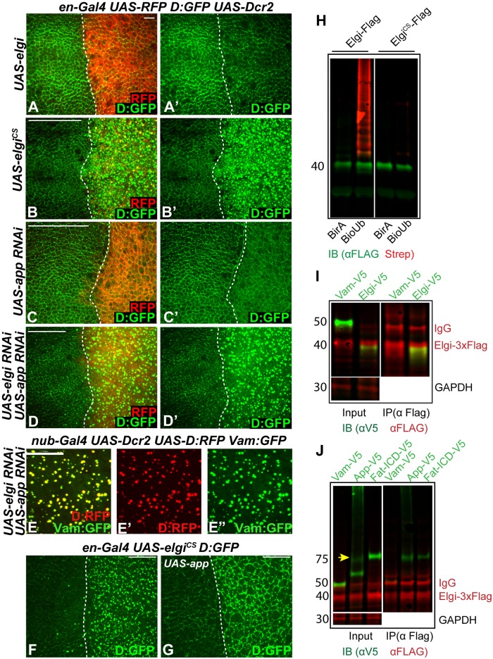
Fig 6. elgiCS has a neomorphic activity and interferes with app function.
(A-D) Horizontal apical sections of wing imaginal discs expressing en-Gal4 UAS-dcr2 D:GFP UAS-RFP along with UAS-elgi-Myc-HA (UAS-elgi) (A, A’), UAS-elgiCS-Myc-HA (UAS-elgiCS) (B, B’), UAS-app-RNAi (C, C’), or UAS-elgi-RNAi and UAS-app-RNAi (D, D’), showing the effect on Dachs:GFP (D:GFP, green) levels and localization in the posterior compartment (marked by RFP, red). Dashed white line marks the A-P compartment boundary. (E-E”) Horizontal apical sections of wing imaginal discs expressing nub-Gal4 UAS-dcr2 UAS-D:RFP UAS-Vam:GFP UAS-elgi-RNAi UAS-app-RNAi showing close up view of D:RFP (red) (E’) and Vam-GFP (green) (E’) colocalization. (F,G) Horizontal apical sections of wing imaginal discs expressing en-Gal4 UAS-elgiCS D:GFP (F) or along with UAS-app (G) showing the effect on localization of Dachs:GFP (D:GFP) in the posterior compartment (to the right). Dashed white line marks the A-P compartment boundary. Scale bar is 6 μm in A, 33 μm in B-D, 17 μm in E and 19 μm in F and G. (H) Western blot showing results of ubiquitination experiments on proteins co-expressed in S2 cells. 3xFLAG tagged Elgi or ElgiCS were co-expressed with either BirA or BirA-BioUb and immunoprecipitated using anti-Flag beads. Elgi was detected by anti-FLAG antibody and biotinylated Ubiquitin was detected with IRDYE680 conjugated streptavidin (Red). (I) Western blot showing results of co-immunoprecipitation experiments on proteins co-expressed in S2 cells. V5-tagged Vam or Elgi were co-expressed with full length Elgi-3x-FLAG, and complexes were precipitated using anti FLAG affinity resin and immunoblotted with anti-V5 and anti-FLAG antibodies. GAPDH was used as a control for loading and transfer in the input lanes. Elgi-3x-FLAG has a slower mobility compared to Elgi-V5 due to the larger epitope tag. (J) Western blot showing results of co-immunoprecipitation experiments on proteins co-expressed in S2 cells. V5-tagged Vam, App (App-V5) or Fat-ICD (Fat-ICD-V5) were co-expressed with Elgi-3x-FLAG, and complexes were precipitated using anti-FLAG affinity resin and then immunoblotted with anti-V5 and anti-FLAG antibodies. GAPDH was used as a control for loading and transfer in the input lanes. Arrow points to the full length App in the input lane.

