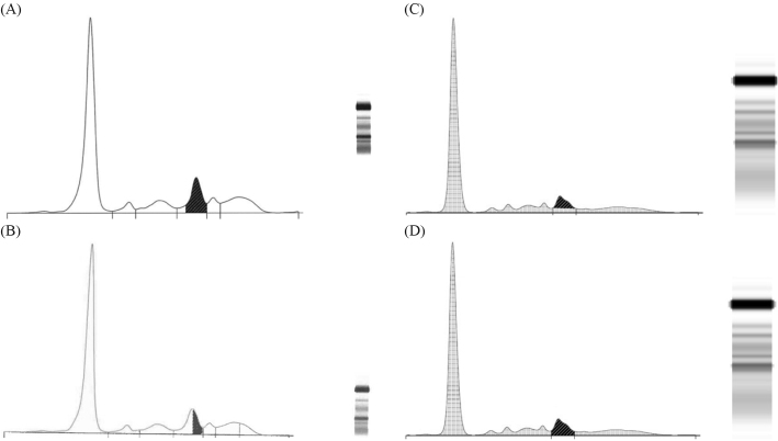
Figure 1.
(A) Densitometric scan of sample 4 analysed by New Zealand (NZ) laboratory 4 using perpendicular drop gating and Capillarys® capillary zone electrophoresis (CZE) methods. IgA lambda was reported as 8 g/L (hatched area). Note the paraprotein is in the beta-1 zone. (B) Densitometric scan of sample 4 analysed by NZ laboratory 3 using corrected perpendicular drop gating and Capillarys® CZE methods. IgA lambda was reported as 4 g/L (hatched area). Note the paraprotein is in the beta-1 zone. (C) Densitometric scan of sample 4 analysed using tangent skimming gating and high-resolution agarose gel electrophoresis (HR AGE) methods. IgA lambda would be reported as 4 g/L (hatched area). Note the paraprotein is in the beta-2 zone. (D) Densitometric scan of sample 4 analysed by NZ laboratory 9 using perpendicular drop gating and HR AGE methods. IgA Lambda was reported as 6 g/L (hatched area). Note the paraprotein is in the beta-2 zone.

