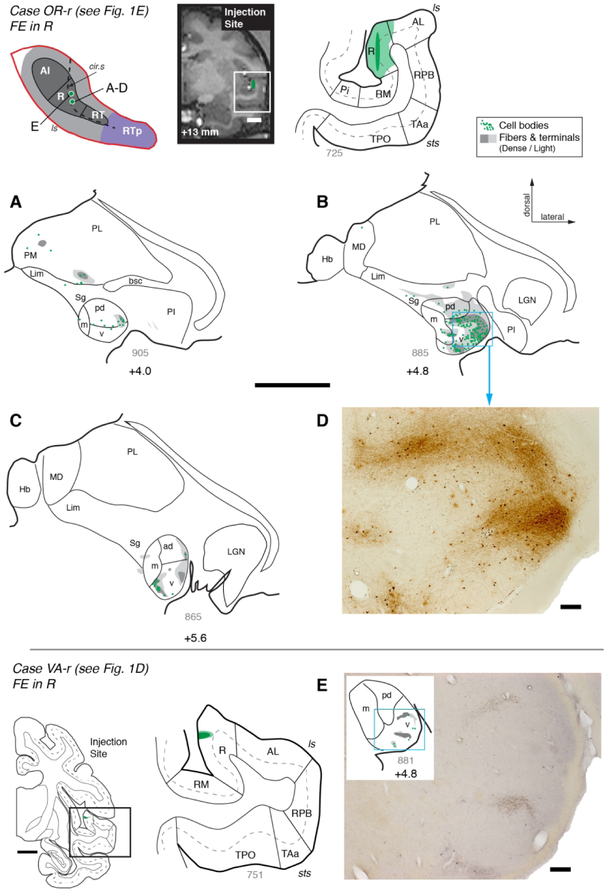
Figure 5.
Thalamic connections of area R in cases OR-r and VA-r. (A-C) Distribution of label after injection of bi-directional tracer FE into area R in case OR-r. Both retrograde and anterograde label were predominantly found within the MGv, where labeled cells were coextensive with dense patches of labeled terminals and fibers. (D) A photomicrograph of the region outlined by the box in panel B shows the FE positive retrograde and anterograde label in the MGv. (E) Results from a smaller injection of FE restricted to the supragranular layers of area R in case VA-r. Scattered cells and a few thin fingers of anterograde label were localized to MGv. Scale bars = 0.2 mm for photomicrographs in D and E, 5 mm for all other panels. For other conventions see Figure 3.

