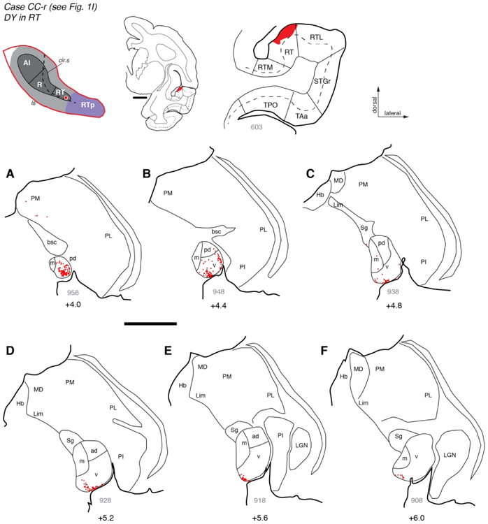
Figure 6.
Thalamic connections of area RT in case CC-r. (A-F) Distribution of labeled neurons after injection of retrograde tracer DY into area RT (see Fig. 1I). Labeled neurons were located primarily in the caudal MGN, including MGpd and MGv. Rostrally, labeled neurons were restricted to the most ventral portion of MGv. The FB injection into RT in another case (Fig. 4) produced a strikingly similar pattern of labeling in the thalamus. Scale bars = 5 mm. For other conventions see Figures 3 and 4.

