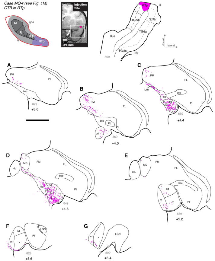
Figure 9.
Thalamic connections of area RTp in case MQ-r. (A-G) Distribution of labeled neurons after injection of retrograde tracer CTB into area RTp, near the border of the STGr (see Fig. 1M). Labeled neurons were located in all subdivisions of the MGN, with a bias toward the posterior half of the nucleus (panels B-D). As was seen after RT injections (Figs. 6 and 8), labeled cells in anterior MGN occupied the ventral edge of MGv and MGm (panels F, G). Relative to RT, a greater proportion of labeled neurons was located outside of the MGN, within the Sg/Lim (panels C, D) and PM (panels A, B). Scale bars = 5 mm. For other conventions see Figure 3.

