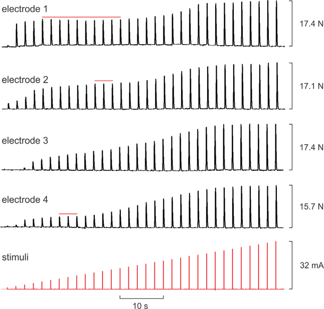
Figure 1.
Example dorsiflexion twitch force responses (upper four traces) to increasing stimulus current pulses (bottom trace) delivered through four intramuscular electrodes placed in different locations within tibialis anterior in one subject. Stimulation through each electrode was performed in separate trials but traces have been aligned for compact display. Red horizontal lines indicate intermediate plateaus wherein force saturated across a range of increasing stimulus intensities.

