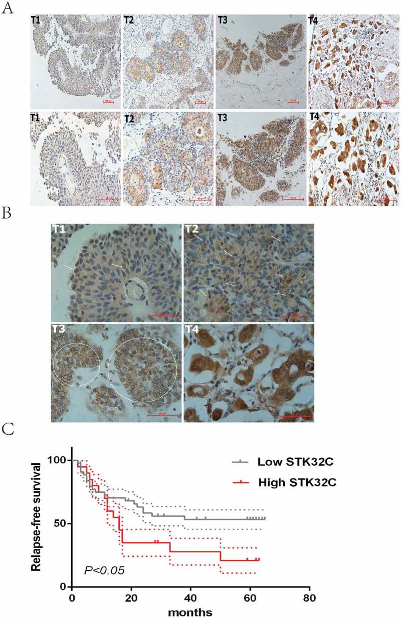
Figure 2.

Immunohistochemistry. (a) Typical staining of STK32C in different stages of BC, a stronger density of staining existed in more invasive BC. (b) Nucleus and cytoplasm of tumor cells was dyed. The arrows and circles referred to the positively stained nucleus. Higher pathologic T stage of BC tended to be more easily stained in the nucleus. (c) Kaplan-Meier analysis was used to analyze the relapse-free survival (RFS) of patients treated by TURBT. High expression of STK32C was associated with a poor RFS with a P < 0.05.
