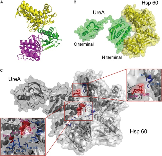
Figure 4.

The interaction analysis between UreA and Hsp60 by molecular docking. (A) Structure of the constructed Hsp60 monomer from H. pylori based on E. coli GroEL (PDB: 2YNJ). (B) The interaction model of UreA (PDB: 1E9Z) and Hsp60 established by molecular docking. (C) Interfaces and key residues analysis. Residues on UreA participating in the interaction are colored in red and noted with purple words. Residues on Hsp60 participating in the interaction are colored and noted in blue.
