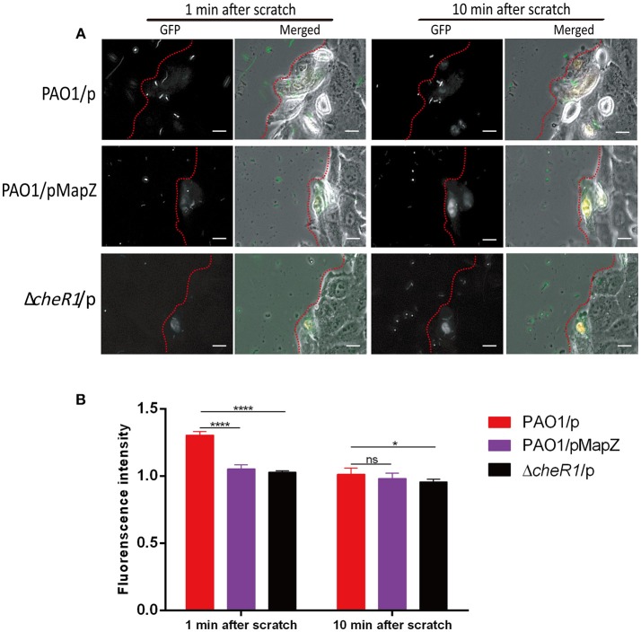
Figure 4.
Chemotaxis-guided migration of P. aeruginosa strains toward scratch-wounded A549 human cells. (A) Representative microscopic images showing the accumulation of P. aeruginosa cells around wounded A549 human cells. The P. aeruginosa cells (in green) and wounded A549 cells (in white) are shown near the edge of the wound as indicated by the red dotted lines. The movies (Movies S1–S3) can be found in the supplementary material. Scale bar = 10 μm. (B) Quantitative comparison of the accumulation of P. aeruginosa cells around the injured A549 cells as indicated by fluorescence intensity. Three independent experiments were performed on each strain and at least 10 cells from each strain were used for quantitative analysis [Data are mean ± SD (n > 10)]. Two-tailed t-test, ****P < 0.0001, *P < 0.05.

