Figure 5.
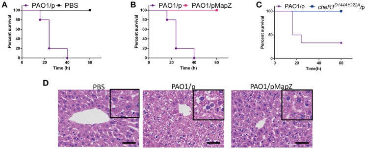
Overexpression of MapZ attenuated P. aeruginosa virulence. Survival rate of mice following intraperitoneal injection of 3.3 × 10⁶ CFU of PAO1/p and PBS (A), 3.3 × 10⁶ CFU of PAO1/p and PAO1/pMapZ strains (B), and 2.4 × 10⁶ CFU of PAO1/p and cheR1D144AY222A strain (C). Data are representative of three independent experiments with five mice used for each group. Histology of liver after intraperitoneal infections. Liver was harvested at the end of 24 h infection and processed for paraffin inclusion. Sections were stained with haematoxylin and eosin staining of liver tissue from the Babl/C mice. All cultures of the strains have the same OD600 at 0.1. (D) Representative photos for haematoxylin and eosin staining of bacteria-infected liver tissue from Babl/C mice after inoculation with PBS, PAO1/p, and PAO1/pMapZ for 24 h, respectively, PBS was used to be a negative control. Tissue infected with PBS can be seen a small amount of liver cells are mild edema around the central vein and edge with cell swelling and cytoplasm loose light dye. Tissue infected with PAO1/p is visible liver cells widely moderate edema with cell swelling and cytoplasm loose light dye, while tissue infected with mapZ_R13A/p, and PAO1/pMapZ only can be seen mild edema liver cells at part of the tissue and edge around the central vein with cell swelling and cytoplasm loose light dye. Data are representative of two independent experiments. Scale bar = 50 μm.

