Figure 5.
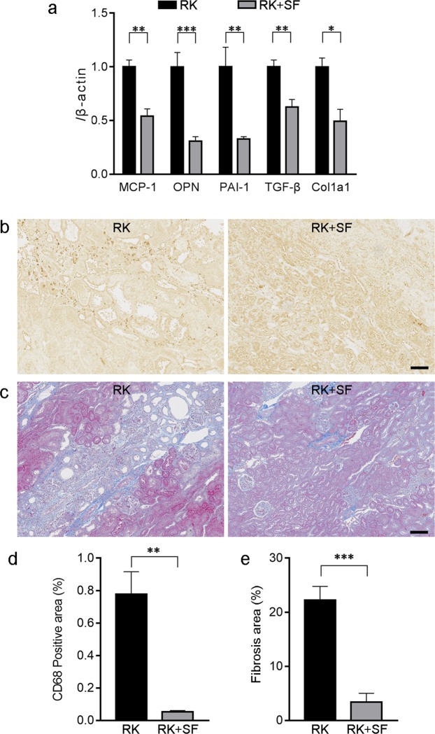
Renoprotective effects of SF is associated with the alleviation of renal inflammation and fibrosis. (a) Quantitative analysis of MCP-1, osteopontin (OPN), PAI-1, TGF-β, and α1(I) collagen (Col1a1) gene expression by real time RT-PCR in the kidney of RK and RK+SF rats (n = 8 each group). (b) Immunohistochemical staining for ED-1 (CD68) in the kidneys of RK (left) and RK + SF (right) rats. Infiltration of the CD68-positive macrophages (indicated by arrows) in RK rats was ameliorated by SF. Bar represents 100 μm. (c) Masson’s trichrome staining of the kidney sections from RK (left) and RK + SF (right) rats. Fibrotic area was reduced by SF administration. Bar represents 100 μm. (d,e) Quantitative analysis of the CD68-positive area (d) and fibrosis area (e) (see also Methods; n = 4 each group). Data are expressed as mean ± SEM; *P < 0.05. **P < 0.01. ***P < 0.001.

