Figure 1.
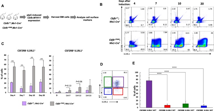
Dysregulated expression of cell surface marker expression by Cbfb-MYH11. (A) Schematic representation of experimental design. Cbfb+/56M; Mx1-Cre+ or Cbfb+/+; Mx1-Cre+ mice were injected with polyinosinic-polycytidylic acid (pIpC) to induce the expression of Cbfb-MYH11. Bone marrow cells were harvested, lineage negative (lin−) cells isolated, and stained with antibodies for IL1RL1 and CSF2RB. (B) Representative FACS plots showing the expression of CSF2RB and IL1RL1 at the indicated time points after the induction of Cbfb-MYH11. (C) Bar graph showing the percentages (%) of CSF2RB− IL1RL1+ (left) and CSF2RB+ IL1RL1+ (right) cells at the indicated time points. (D) Representative IL1RL1 and KIT staining of CSF2RB− cells from leukemic Cbfb-MYH11+/56M, Mx1-Cre+ mice. (E) Bar graph of IL1RL1 and KIT expression in CSF2RB− population. N ≥ 3; **P < 0.01; ****P < 0.0001.

