Fig. 5.
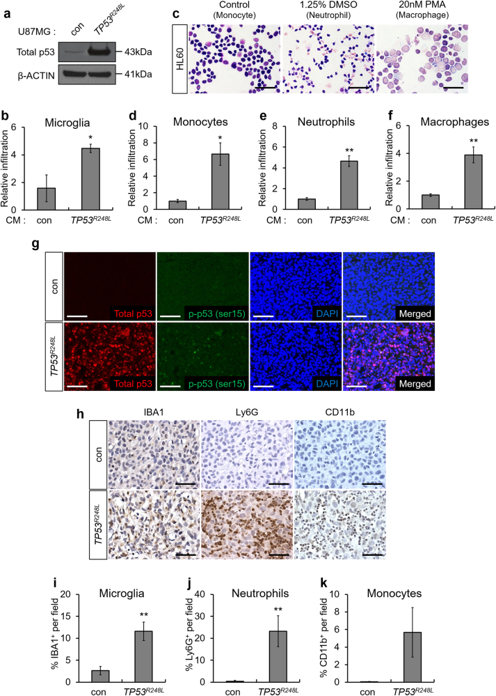
Ectopic expression of TP53R248L promotes immune cell infiltration. a Western blot analysis showing p53 expression in TP53R248L-overexpressing U87MG cell line. b Relative infiltration rate of BV2 microglia grown in conditioned medium (CM) generated using control U87MG and TP53R248L-overexpressing U87MG cell lines (n = 3). c Differentiation of HL60 to neutrophils and macrophages was confirmed by Diff-Quik staining (magnification 400×, scale bar = 50 μm). d–f Relative infiltration rate of HL60-derived monocytes, neutrophils, and macrophages grown in CM from control U87MG and TP53R248L-overexpressing U87MG cell lines (n = 3). g Representative microscopic images of fluorescent IHC showing total p53 and p-p53 (ser15) in orthotopically transplanted tumors generated using TP53R248L-overexpressing U87MG cell line (magnification 200×, scale bar = 100 μm). h Representative microscopic images of IHC showing expression of immune cell markers such as IBA1, Ly6G, and CD11b (magnification 200×, scale bar = 100 μm). i–k Quantification of IHC showing the composition of IBA1+, Ly6G+, and CD11b+ cells. The bar graphs represent mean ± SEM (*P < 0.05; **P < 0.01; ***P < 0.001)

