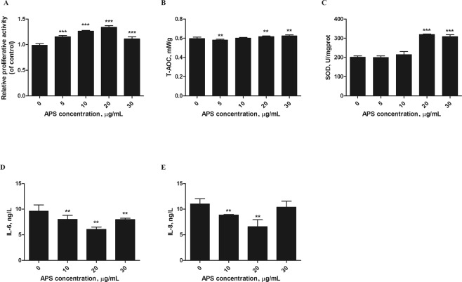
Figure 2.
APS protected MEFs against H2O2-induced cell damage. MEFs were stimulated with 250 μM H2O2 for 12 h, and then incubated with different APS concentrations (0–30 µg/mL). (A) Cell proliferation was then measured using the MTT method. (B,C) The T-AOC and SOD activity in the cells were analyzed at 450 nm. (D,E) The concentrations of IL-6 and IL-8 were detected by ELISA. The values represent the mean ± SD (n = 3 per group); **P < 0.01 and ***P < 0.001.

