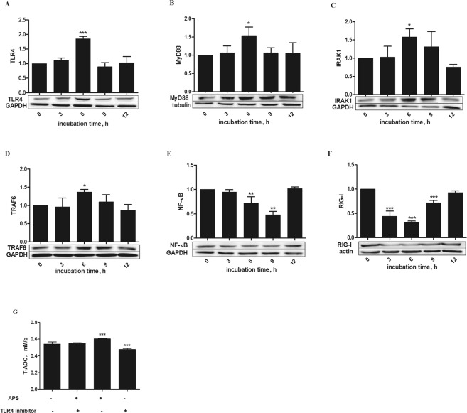
Figure 3.
Effects of APS on expression of key proteins in the TLR4 signaling pathway, NF-κB and RIG-I in H2O2-pretreated MEFs. (A–F) Cells were treated for varying times (0, 3, 6, 9 and 12 h) with 20 µg/mL APS after stimulation with 250 µM H2O2 for 12 h. Proteins levels were determined by Western blotting, and the band intensities were analyzed using image analysis software. (G) Cells were stimulated with 250 µM H2O2 for 12 h. The MEFs were incubated in the presence or absence of a TLR4-neutralizing antibody (20 μg/mL) for 1 h and then treated with 20 μg/mL APS for 6 h, and the T-AOC was determined. The data are presented as the mean ± SD (n = 3 per group), and significance relative to the control group was determined using Duncan’s multiple-range test; *P < 0.05 and ***P < 0.001.

