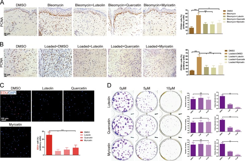
Fig. 3. Luteolin, quercetin or myricetin inhibits fibroblast proliferation in vivo and in vitro.
Representative images of hypertrophic scar (HS) in a fibrosing skin in bleomycin-induced model and b mechanical load-induced HS model from DMSO-treated mice and drug-treated mice stained for proliferating cell nuclear antigen (PCNA) and quantitative analysis of PCNA-positive cells. c Representative immunofluorescence images and quantitative results of EdU incorporation assay in human dermal fibroblasts (HDFs). EdU is shown by red fluorescence and nucleus by blue fluorescence. d Representative images and quantitative results of colony formation assay exhibit an evidently reduced number of HDF colony (purple) after drug treatment. Data are the mean ± SD (three independent experiments); *P < 0.05, ***P < 0.001

