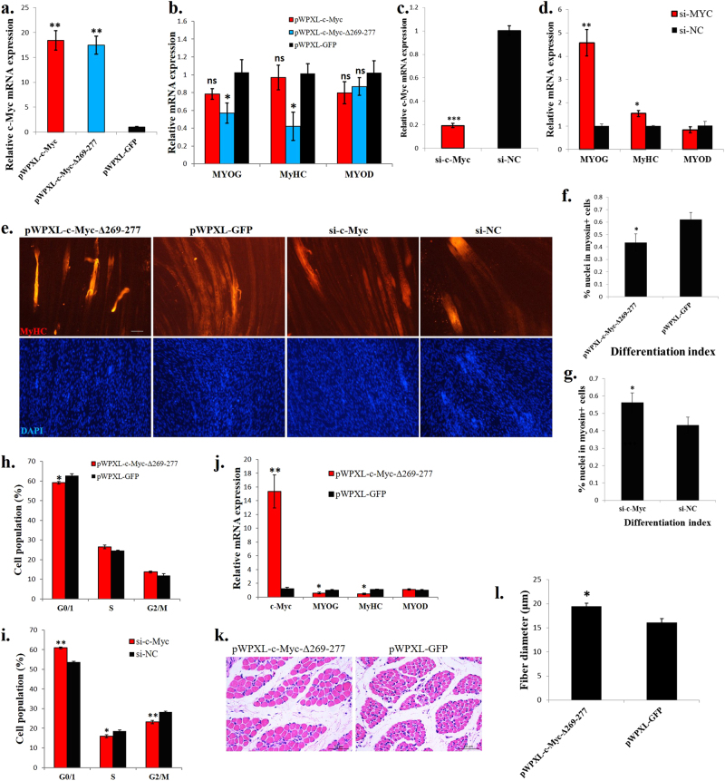
Fig. 2.
c-Myc regulates myoblast proliferation and differentiation in vitro and induces muscle fibre hypertrophy in vivo. a Relative c-Myc mRNA expression after introducing c-Myc, c-Myc-Δ269–277 and GFP into chicken primary myoblasts. b Muscle differentiation marker genes expression after introducing c-Myc, c-Myc-Δ269–277 and GFP into chicken primary myoblasts. c Relative c-Myc mRNA expression after c-Myc knockdown in chicken primary myoblasts. d Muscle differentiation marker gene expression after c-Myc knockdown in chicken primary myoblasts. e MyHC immunostaining of primary myoblasts transduced with indicated vectors or siRNAs. Cells were differentiated for 72 h after transfection. The nuclei were visualized with DAPI. Bar, 100 µm. f Differentiation index of cells expressing c-Myc-Δ269–277 or GFP. g Differentiation index of cells transfected with si-c-Myc or si-NC. h Primary myoblasts expressing c-Myc-Δ269–277 or GFP were cultured in GM, and the cell cycle phase was analysed after 2 days. i Primary myoblasts transfected with si-c-Myc and si-NC were cultured in GM, and the cell cycle phase was analysed after 2 days. j Relative mRNA expression of the indicated genes in chicken breast muscles infected with a c-Myc-Δ269–277-expressing lentivirus or control (GFP). k H–E staining of a breast muscle fibre cross section from chickens infected with a c-Myc-Δ269–277-expressing lentivirus or control (GFP). l Fibre diameter of chicken breast muscle infected with a c-Myc-Δ269–277-expressing lentivirus or control (GFP). The results are shown as the mean ± sem of three independent experiments. In a, b, ANOVA followed by Dunnett’s test was used. In c, d, f–j, l, independent sample t-test was used. *p < 0.05; **p < 0.01; ns, no significant difference

