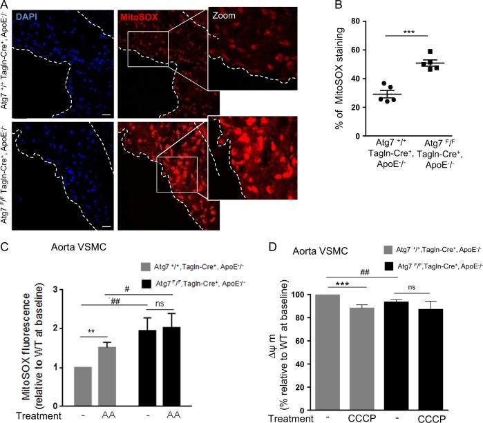
Fig. 3. Increased mitochondrial reactive oxygen species (ROS) production and reduced mitochondrial membrane potential in plaques from ApoE−/− mice deleted for Atg7 in vascular smooth muscle cells (VSMCs).
a Representative images of consecutive aortic sinus sections stained with MitoSOX (red) and DAPI (blue, nucleus) of Atg7+/+ Tagln-Cre+, ApoE−/− and Atg7F/F Tagln-Cre+, ApoE−/− mice after 10 weeks of high-fat diet (HFD). Scale bar, 20 µm. b The graph represents the % of MitoSox staining in the plaque area per section and the data are the mean ± SEM. ***P < 0.001; Student’s t-test, n = 5 mice/group. c Measurement of mitochondrial ROS production in VSMCs isolated from the aorta of Atg7+/+ Tagln-Cre+, ApoE−/− and Atg7F/F Tagln-Cre+, ApoE−/− mice after 10 weeks of HFD. The graph represents the quantification of MitoSOX Red fluorescence at baseline or after antimycin A (AA, 10 µM) stimulation. Data are the median with interquartile range of four independent experiments from different primary VSMC cultures per group. **P < 0.01; ##P < 0.01; #P < 0.05; ns, nonsignificant; one-way ANOVA, Kruskal–Wallis non-parametric test. d Measurement of the mitochondrial membrane potential (ΔΨm) with the JC-1 dye in VSMCs isolated from the aorta of Atg7+/+ Tagln-Cre+, ApoE−/− and Atg7F/F Tagln-Cre+, ApoE−/− mice after 10 weeks of HFD. The graph represents the quantification of the potential-dependent accumulation of the JC-1 dye in mitochondria at baseline and after CCCP (20 µM) stimulation. Data are the median with interquartile range of four independent experiments from different primary VSMC cultures per group. ***P < 0.001; ##P < 0.01; ns, nonsignificant; one-way ANOVA, Kruskal–Wallis non-parametric test

