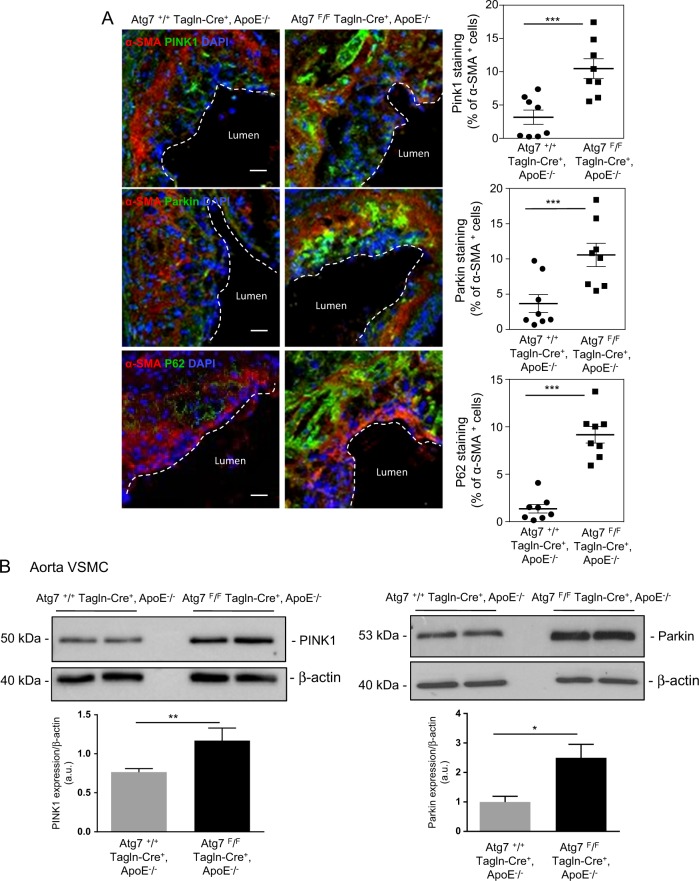
Fig. 5. Increased expression of the mitophagy proteins PTEN-induced putative kinase 1 (PINK1) and Parkin functions in VSMCs from atherosclerotic lesions of ApoE−/− mice deleted for Atg7 in vascular smooth muscle cells (VSMCs).
a Representative images of consecutive aortic sinus sections immunostained with PINK1, Parkin, P62 (red), α-SMA (green) antibodies, and DAPI (blue, nucleus) from Atg7+/+ Tagln-Cre+, ApoE−/− and Atg7F/F Tagln-Cre+, ApoE−/− mice after 10 weeks of high-fat diet (HFD). The graph represents the % of PINK1, Parkin, or P62 staining in VSMCs within the plaque area and the data are the mean ± SEM from n = 8 mice/group. ***P < 0.001; Student’s t-test. Scale bar, 20 µm. b Western blot analyses of the expression of PINK1 and Parkin proteins in aortic VSMCs isolated from Atg7+/+ Tagln-Cre+, ApoE−/− and Atg7F/F Tagln-Cre+, ApoE−/− mice after 10 weeks of HFD, β-actin was used as the loading control. Bands are shown in duplicate. The graph represents the densitometric analysis of the expression level of PINK1 and Parkin proteins. The data are the mean ± SEM of three independent experiments from different primary VSMC cultures per group. **P < 0.01; *P < 0.05; Student’s t-test

