Fig. 2.
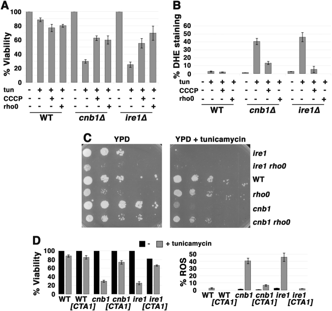
ER stress-induced death is caused by mitochondrial ROS. a ER stress-induced death is rescued in rho0 cells and by addition of the proton ionophore CCCP. Exponentially growing cells were treated with 0.5 µg/ml tunicamycin with or without CCCP (10 µM) for 5 h. Cell viability was then determined as in Fig. 1b legend, expressed as a percentage of untreated cells +/− SEM, n = 3. b ROS accumulation is rescued in rho0 cells and by addition of the protonophore CCCP. ROS was assayed by staining with DHE as in Fig. 1c legend; n = 3+/− SEM. Fluorescence images from cells +/− tunicamycin were imaged for the same exposure time and adjusted using identical Photoshop settings. c Impaired growth of cnb1 on tunicamycin is rescued in cnb1 rho0 cells. Serially diluted cells were spotted onto plates with and without tunicamycin (0.25 µg/mL). d ER stress-induced death (left panel) and ROS (right panel) rescued by overexpression of mitochondrial catalase. Cells transformed with pMET-CTA1 were induced to express catalase after washing with water and resuspending in methionine-free SC medium. Tunicamycin (0.5 µg/mL) was then added for 5 h before measuring cell viability and ROS as in Fig. 1b, c legends +/− SEM, n = 3.

