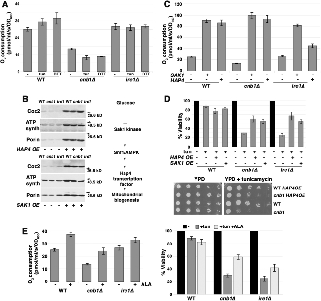
Fig. 3.
ER stress induces mitochondrial response. a O2 consumption increases in response to ER stress in wild-type but not cnb1∆ or ire1∆ cells. Cells were treated with 0.5 µg/mL tunicamycin or 1 mM DTT for 2 h before measuring O2 consumption expressed as pmol/ml/s/OD600+/− SEM, n = 3. b ETC protein levels are increased by overexpression of HAP4 (top left panel) and SAK1 overexpression (bottom left panel). Lysates from exponentially growing wild-type and cnb1∆ cells +/− HAP4 overexpression were assayed by Western blot for ETC components, Cox2 and ATP synthase, and mitochondrial outer membrane protein VDAC1/porin. Right panel, schematic indicating signaling pathways regulating mitochondrial biogenesis. c O2 consumption is increased upon overexpression of HAP4 or SAK1. O2 consumption is increased in cells overexpressing HAP4 or SAK1. d Overexpression of HAP4 or SAK1 suppresses death of cnb1∆ and ire1∆ cells after 5 h tunicamycin treatment. Top panel, cell viability was assayed as in Fig. 1b legend. Bottom panel, HAP4 overexpression relieves impaired growth of cnb1∆ cells on tunicamycin. Serial dilutions of cells were spotted on SC-uracil plates with 0.2 µg/mL tunicamycin. e ALA to promote heme synthesis increases O2 consumption (left panel) and rescues viability (right panel) of ire1∆ and cnb1∆ cells during ER stress. Cells were grown overnight in SC medium supplemented with ALA to mid-log phase. Viability was determined after 5 h treatment with tunicamycin (0.5 µg/mL) as described in Fig. 1b legend.

