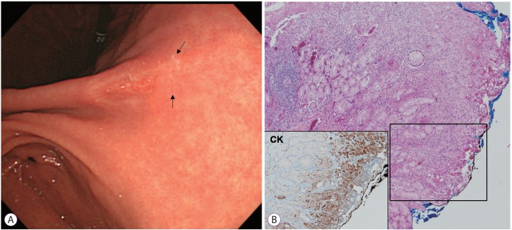
Fig. 1.
Signet ring cell carcinoma (SRC) case with positive lateral margin after endoscopic resection. (A) Endoscopic image of early gastric cancer, showing a depressed lesion located in the posterior wall of the angle. The surrounding mucosa was combined with atrophic gastritis. After endoscopic resection, the lateral margin was positive (arrow). (B) Pathological findings after endoscopic resection (hematoxylin and eosin, ×40). SRC cells showed subepithelial spread. Immunohistochemical staining for CK (AE1/AE3), ×100.

