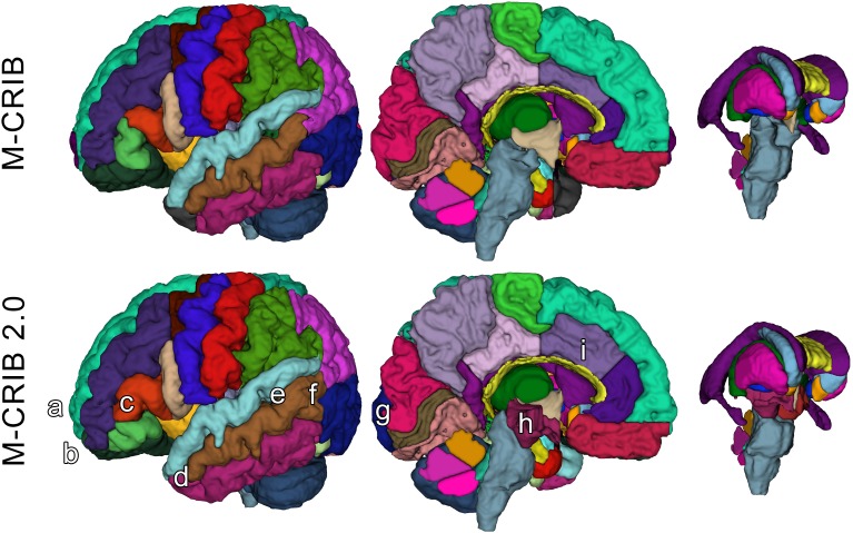
FIGURE 1.
Surface meshes of a single left hemisphere (lateral and medial views) and some subcortical structures, of a single participant, illustrating some examples of updated regions. Top row: original M-CRIB atlas. Bottom row: M-CRIB 2.0. Annotations indicate some of the updates made: (a) removal of frontal pole, (b) revision of boundary between lateral orbitofrontal (dark green) and rostral middle frontal (dark blue) regions, (c) revision of boundaries of ‘pars’ regions of inferior frontal gyrus. (d) Replacement of temporal pole (dark gray) with superior, middle, and inferior temporal labels, (e) replacement of ‘banks STS’ (dark green) region with superior and middle temporal labels, (f) revision of boundary between lateral occipital (dark purple) and temporal regions, (g) revision of medial boundary of lateral occipital region, (h) addition of ‘ventral diencephalon’ (maroon) which replaces sections of brainstem (gray) and removed ‘subcortical matter’ (not shown) label, (i) revision of rostral, and caudal anterior cingulate (purple) regions to encompass cortex extending to the more rostral/dorsal branch of a parallel double cingulate sulcus.

