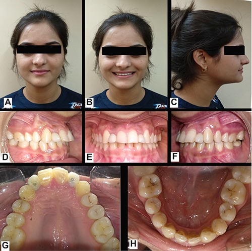
Figure 2.

Post-treatment photographs. (A) extraoral front view; (B) extraoral smiling view; (C) extraoral profile view; (D) right lateral view of dentition; (E) in occlusion front view of dentition; (F) left lateral view of dentition; (G) occlusal view of maxillary arch; (H) occlusal view of mandibular arch.
