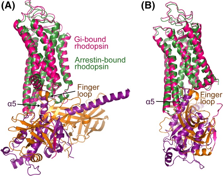
Figure 7.

Comparison of the interfaces of Gαi and visual arrestin with rhodopsin. (A) Gαi α5 can be closely superpositioned with the arrestin finger loop when Gi‐bound rhodopsin is aligned with arrestin‐bound rhodopsin. Gi‐bound rhodopsin is colored in red, arrestin‐bound rhodopsin in dark green, Gαi in magenta, and visual arrestin in brown. (B) A view with 90° rotation about the vertical axis.
