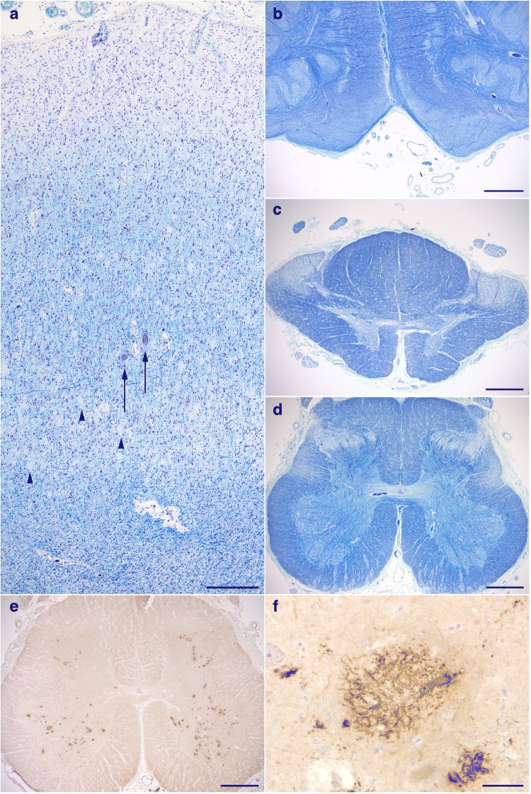
Fig. 6.
Degeneration of upper motor neurons. a Loss of Betz cells. (Arrows) A few remaining Betz cells. (Arrowheads) Numerous unstained CWPs that tend to be densely distributed in the deep cortical layers. b Degeneration with evident atrophy of the pyramidal tract at the level of the medulla oblongata. c, d Degeneration in the lateral tract in the thoracic (c) and lumbar cords (d). The anterior horn cells are well preserved in number (d). e Remarkable Aβ deposits in the anterior horns in the lumbar cord, while no Aβ deposit is seen in the corticospinal tract. f A high power view of Aβ deposits in the anterior horn in the lumbar cord. Scale bars = a 300 μm, b-e 1 mm, f 50 μm. a-d Klüver-Barrera stain. e, f 12B2 immunohistochemistry

