Fig. 3.
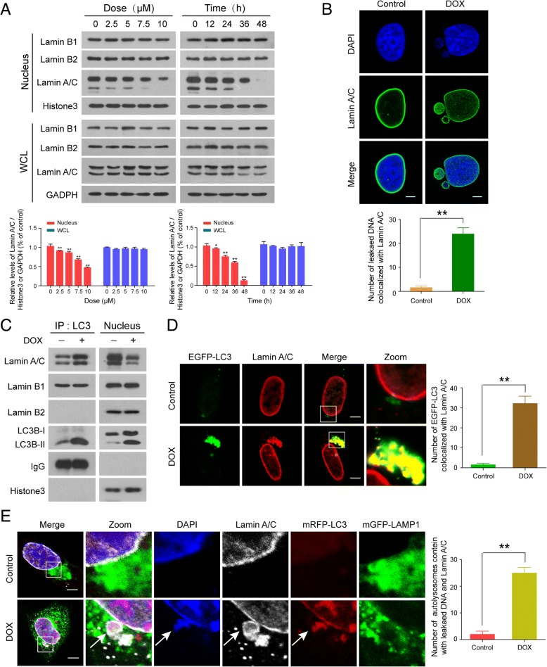
LC3-lamin A/C interaction is required for nucleophagy. MDA-MB-231 cells were treated without or with DOX as indicated. (a) The expressions of nuclear proteins, lamin A/C, lamin B1, and lamin B2, in whole cellular (WCL) and nuclear extracts (nucleus) were detected by western blot. Comparison of the intensities were statistically estimated and represented as mean ± SD for three independent experiments (*P < 0.05, **P < 0.01). (b) The colocalization of lamin A/C (green) and leaked nuclear DNA (blue) was determined by confocal microscopy. Scale bars: 10 μm. The number of DAPI-stained particles with leaked nuclear DNA colocalized with lamin A/C was quantified from 30 cells in three independent experiments. Data was presented as mean ± S.D. (**P < 0.01). (c) After treatment with DOX (10 μM, 24 h), nuclear lysates were prepared and subjected to immunoprecipitation using anti-LC3, and the associated LC3B-I/LC3B-II, lamin A/C, lamin B1, and lamin B2 were determined by immunoblotting. (d) MDA-MB-231 cells were transfected with EGFP-LC3, treated as in (b), and examined by confocal microscopy to determine the colocalization of EGFP-LC3 and lamin A/C (red). Scale bars: 10 μm. The number of EGFP-LC3 colocalized with lamin A/C was quantified from 30 cells in three independent experiments. Data was presented as mean ± S.D. (**P < 0.01). (e) Immunofluorescence analysis showed the colocalization of leaked DNA (blue) with mRFP-LC3(red), mGFP-LAMP1(green), and lamin A/C (white) in MDA-MB-231 cells treated with DOX (10 μM, 24 h). Scale bars: 10 μm. The number of autolysosomes containing leaked DNA and lamin A/C was quantified from 50 cells in three independent experiments. Data was presented as mean ± S.D. (**P < 0.01)

