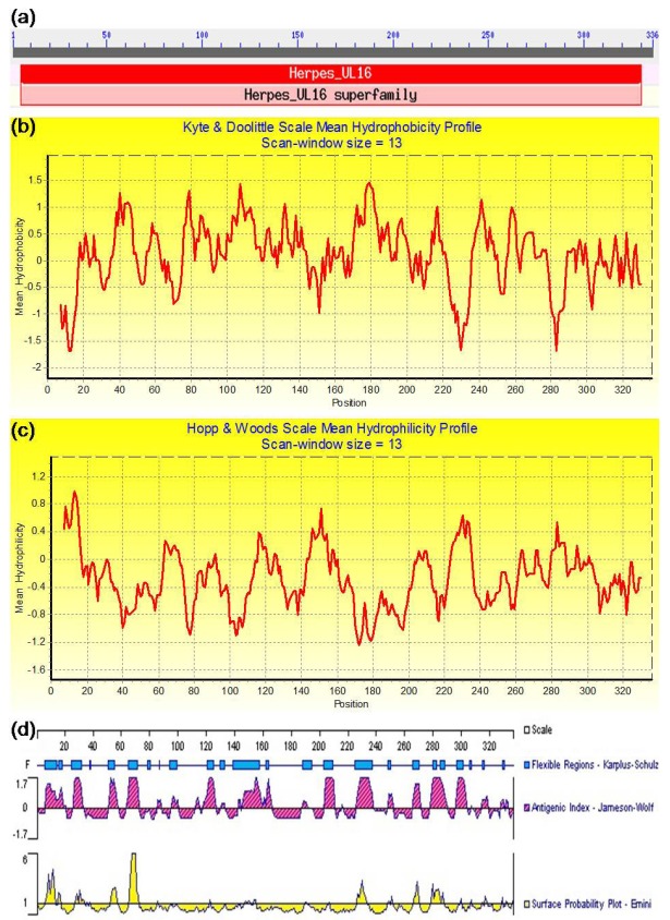
Figure 7.
The conserved domain, hydrophobicity, hydrophilicity, and antigenic analyses of the EBV BGLF2. (a) The conserved domain analysis of the BGLF2 using NCBI Conserved Domains search tool. (b) The hydrophobicity or (c) hydrophilicity profile was determined using the values of Kyte and Doolittle (Kyte and Doolittle, 1982) or Hopp and Woods (Hopp and Woods, 1981), respectively, with a 13-amino-acid window. The peaks pointing up represent the most hydrophobic (b) and hydrophilic (c) regions, respectively. (d) Antigenic analysis of the BGLF2 was carried out through determination of its primary structure using the PROTEAN software of the DNAStar based on its flexibility, antigenic index, and surface probability.

