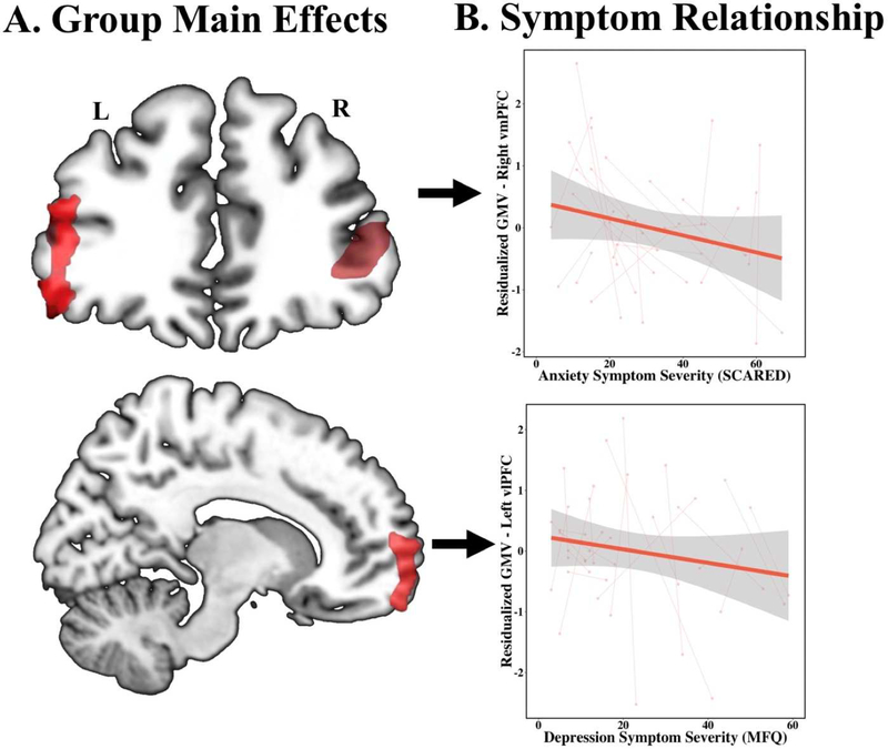
Figure 1: Gray matter abnormalities and symptom correlations in pediatric PTSD (n=22) as compared to TD youth (n=20).
(A) Reduced grey matter volume in the bilateral vlPFC and the right vmPFC in youth with PTSD as compared to TD youth (n=20), covaried for age at baseline, sex, total intracranial volume, and subject as a random effect. (B) Depression symptoms of PTSD, as measured by the MFQ, are inversely correlated with bilateral vlPFC gray matter volume and anxiety symptoms, as measured by the SCARED, are inversely correlated with vmPFC gray matter volume, covaried for age at baseline, sex, total intracranial volume, subject as a random effect, and the four alternative symptom scores (PTSD-RI cluster B, C, D, and SCARED). Scatterplot shows extracted cluster data in relation to depression symptom severity.
Abbreviations: PTSD, post-traumatic stress disorder; TD, typically developing; vlPFC, ventrolateral prefrontal cortex; MFQ, Mood and Feelings Questionnaire; vmPFC, ventromedial prefrontal cortex; SCARED, Screen for Child Anxiety Related Disorders.

