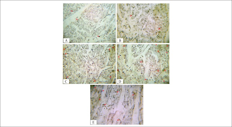
Figure 3.
Immunohistochemical detection of CD31 in myocardial vessels of different groups. Brown stained tissues show CD-31 immunostained endothelial cells in: (A) Control; (B) Diabetes; (C) Diabetes+Garlic; (D) Diabetes+Exercise; and (E) Diabetes+Garlic+Exercise. The intensity of immunostaining for CD31 (arrow head) decreased in the diabetes group compared to the control group. Garlic treatment and exercise alone or combined increased angiogenesis in diabetes compared to the diabetes group (Magnification was 400x).

