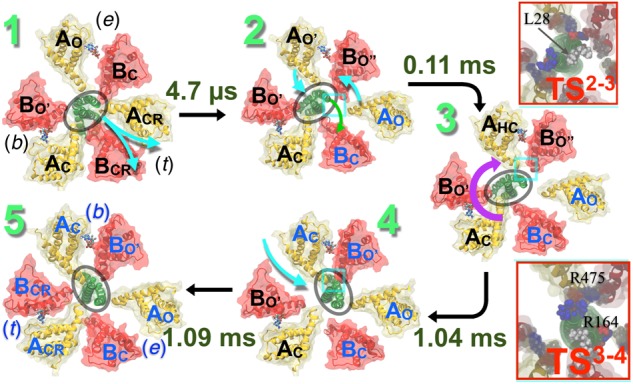
FIGURE 6.

Conformational transitions mechanism of V1 proposed by computer simulation studies. Pathway of the hydrolysis-driven conformational transition in the entire V1 domain derived from the string method with swarms of trajectories (Pan et al., 2008). Firstly, the transition from a ‘tight’ (t) to an ‘empty’ (e) conformation (1→2) is observed at the ADP+Pi-bound site, where ACRBCR is transformed to AOBC. This transition promotes ATP binding at the neighboring ‘empty’-site. This empty site transforms from the AOBC conformation in state 1 to a bindable AO′BO″ form in state 2. ATP-binding to the bindable site yields the first doubly bound state (2→3); the ATP binding simultaneously induces a local deformation of the DF-shaft. The shaft then rotates yielding the second doubly bound state (3→4). Finally, a bound ATP (b) in the third site, in ACBO′ form, transitions to the reaction mode in the ‘tight’ (t) conformation, promoting subsequent hydrolysis (4→5). Rates of each of these transitions are computed employing techniques described in Singharoy and Chipot (2017) and Singharoy et al. (2017) and labeled along with each step. The rotation step is found to be the slowest when it follows product release. (Upper inset) The 2→3 transition necessitates a wring deformation of the shaft that marginally exposes the hydrophobic residue L28 to water. This unfavorable solvation characterizes the first TS (TS23). (Lower inset) Salt bridges between residues of the shaft and those of the AHC and BO″ subunits reorganize during the shaft-rotation step, involving transient repulsive electrostatic interactions of residues R164 (DF) with R475 (AHC). These repulsive interactions characterize the second TS (TS34), featuring the highest barrier of the V1–rotation pathway. Blue beads indicate basic residues, red indicates acidic and white indicates hydrophobic ones. Cumulative transition times are recorded at each step: transition 1→2 takes 4.7 μs, 1→3 takes 0.11 ms, 1→4 takes 1.04 ms, and altogether transitions 1→5 takes 1.09 ms. Figure adapted from Singharoy et al. (2017).
