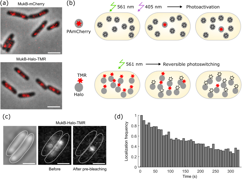
Figure 3.
Microscopy data acquisition using Halo tag. (a) Snapshots of MukB-Halo-TMR and MukB-mCherry recorded with identical settings on an epifluorescence microscope (red: fluorescence with 150 ms exposure, grey: phase contrast). Scale bars 2 µm. (b) Principle of data acquisition for single-molecule localization microscopy using PAmCherry or Halo-TMR labels. (c) Snapshots showing MukB-Halo-TMR intensity before and after pre-bleaching (15 ms exposure). Scale bars 1 µm. (d) Frequency of MukB-Halo-TMR localizations over the course of a movie at constant 561 nm excitation (0.2 kW cm−2), normalised to the initial frequency.

