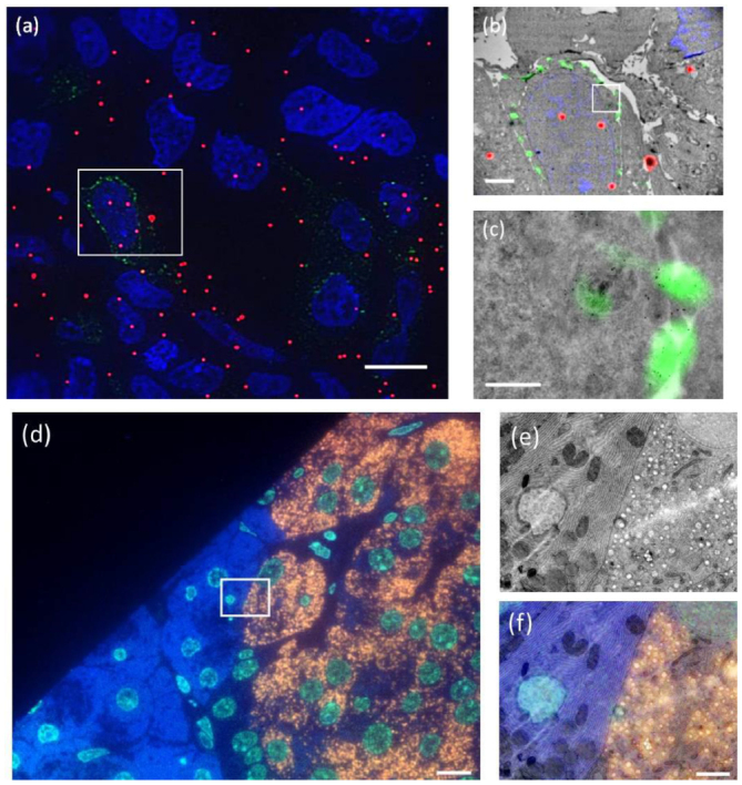
Figure 2.

Examples of (a)–(c) fiducial and (d)–(f) non-fiducial based image registration in integrated microscopes. (a) FM image in TEM (implementation according to figure 1(d)) of Tokuyasu sections of HeLa cells transfected with LAMP-1-GFP. Nuclei are shown in blue (DAPI), LAMP-1-GFP in green and fiducials in red. (b) Overlay of ROI (boxed area in (a)) of fluorescence and TEM images. (c) Zoom in on LAMP-1-GFP rich area. Fiducials consist of silica particles with a 15 nm gold core and a 40 nm fluorescently labeled silica shell. Overlay accuracy is about 30 nm. (d) FM image in SEM (implementation according to figure 1(a)) of rat pancreas sections, immuno-labelled after embedding in epon to show nuclei in blue (Hoechst), guanine quadruplexes in light blue (Alexa488), and insulin in orange (Alexa594). (e) SEM image of the ROI (boxed area in (d)). (f) Overlay of fluorescence from the ROI with the SEM image. The overlay (<20 nm accuracy) is obtained via an automated registration procedure between both microscopes [10]. Scale bars are 10 µm in (a), (d), 2 µm in (b), (e) and (f), and 0.5 µm in (c).
