
Figure 7.
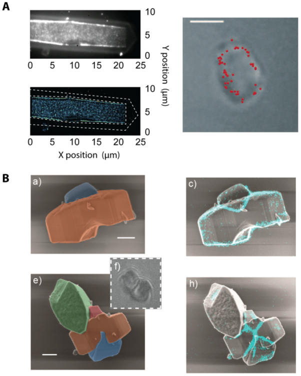
(A) When studying catalyst materials, optical microscopy can be used to visualize chemical events, e.g. using fluorogenic reagents (top left). By carefully tuning reaction conditions, the locations of chemical events can even be mapped on a particle with nanometer accuracy (bottom and right left). Unfortunately, the diffraction limit allows only a very limited amount to be derived from the particle itself (right), making it hard to correlate chemical reactivity with nanoscale structural features. Reproduced from [42] with permission of The Royal Society of Chemistry. (B) The combination of SRF imaging and EM allows individual chemical events to be correlated with ultrastructure at the single particle level. Reprinted with permission from [48]. Copyright 2017 American Chemical Society.