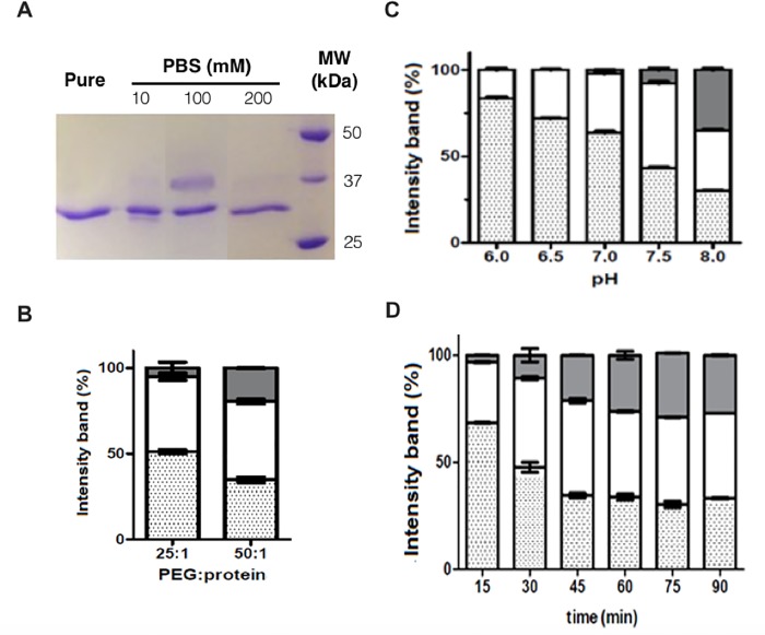
Fig 1. Reaction conditions to produce monoPEGylated L-asparaginase (monoPEG-ASNase).
(A) Electrophoresis (SDS-PAGE) showing the influence of PBS buffer ionic strength on N-terminal PEGylation with pH 7.5 (PEG:ASNase ratio of 25:1, 2 kDa mPEG-NHS): Column 1- ASNase (control), column 2- reaction in 10 mM PBS, column 3- reaction in 100 mM of PBS, column 4- reaction in 200 mM PBS, and column 5- Molecular weight (BioRad). (B) Percentage of PEGylation at different PEG:ASNase ratios, 25:1 and 50:1, in 100 mM of PBS pH 7.5, 30 min of time reaction. (C) Percentage of PEGylation at different pH values (6.0, 6.5, 7.0, 7.5 or 8.0) in 100 mM of PBS, PEG:ASNase ratio of 25:1 and 30 min of reaction time. (D) Percentage of PEGylation on different reaction times (15 to 90 minutes) in 100 mM of PBS, pH 7.5, PEG:ASNase ratio of 25:1. Grey bars—polypegylated ASNase, white bars—monoPEGylated ASNase, dotted bars—free ASNase. Percentage of PEGylation was based on gel analysis by band intensity.

