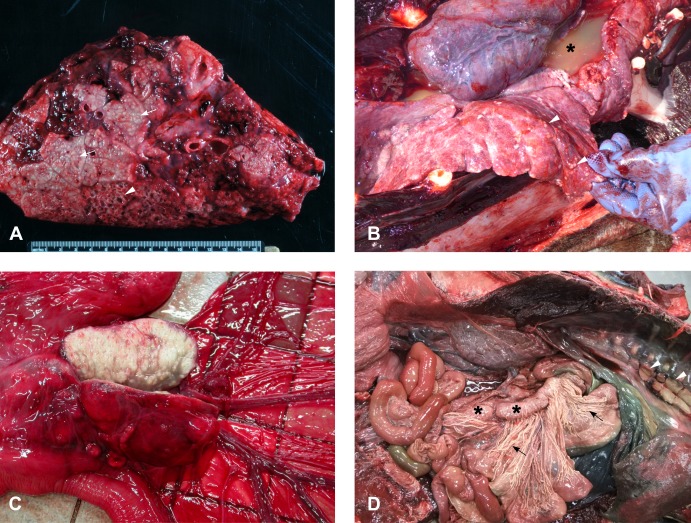
Fig 1. Gross pathology of M. pinnipedii infection.
A. Cut surface of lung from a severely affected New Zealand sea lion. There are areas of lung consolidation containing multifocal to coalescing caseating granulomas (arrows). In some areas these have central areas of liquefaction and tissue loss (arrowhead). B. Miliary lesions: there are numerous small (2-4mm diameter) granulomatous lesions (arrowheads) scattered throughout the lung fields of this New Zealand sea lion, along with watery turbid fluid within the thoracic cavity (pleural effusion; asterisk). C. Cut surface of an enlarged, pale, firm, nodular mesenteric lymph node from a New Zealand sea lion with tuberculosis. D. Tuberculosis in a Hector’s dolphin. The mesenteric (asterisk) and intra-thoracic (arrowheads) lymph nodes are enlarged and pale and the mesenteric lymphatics are pale and thickened (arrows).

