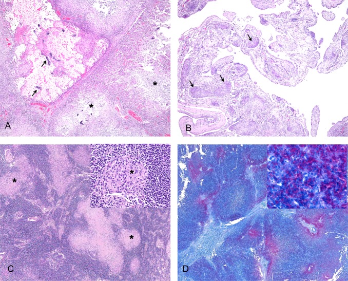
Fig 2. Histopathological lesions present in marine mammals diagnosed with tuberculosis due to Mycobacterium pinnipedii.
A. Lung of a sea lion showing focally extensive destruction of normal parenchyma (top left corner) with mineralisation of alveolar septae. Adjacent tissue shows multifocal granulomas with central areas of necrosis (asterisks), 4x, H&E. B. Mediastinal tissue from a sea lion, which is thickened by diffuse granulomatous inflammation and multiple small granulomas (arrows), 4x, H&E. C. Lymph node from a New Zealand sea lion, showing multifocal to coalescing granulomas (asterisks), 4x. Inset shows a single small granuloma (asterisk) surrounded by normal lymphoid tissue, 40x. Both stained with H&E. D. Superficial lymph node from a Hector’s dolphin. Areas of necrosis are outlined by large numbers of red-stained acid-fast bacilli, 4x. Inset shows high power magnifaction of acid-fast bacilli, 100x. Both stained with Ziehl-Neelsen stain.

