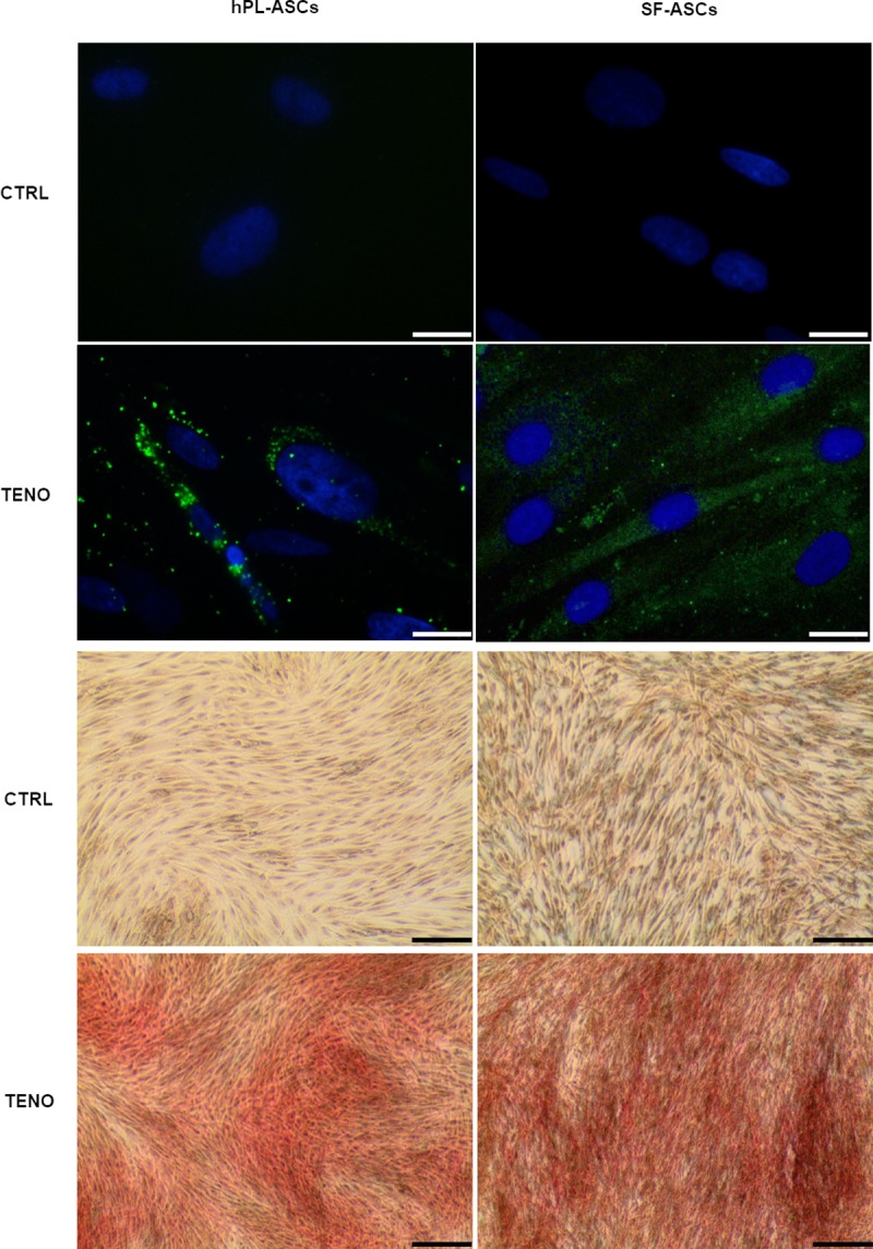
Fig 8. Scleraxis expression and collagen matrix deposition after tenogenic differentiation.

Upper four panels show representative images of scleraxis expression (green) in CTRL and TENO cells at 3 days of differentiation (the nuclei were stained with DAPI, blue) captured by fluorescence microscope (40x; scale bar 20 μm); four panels on the button show representative images related to the collagen matrix deposition, stained by Sirius Red (10x; scale bar 100 μm), that occurred after 7 days differentiation in hPL-ASCs and SF-ASCs.
