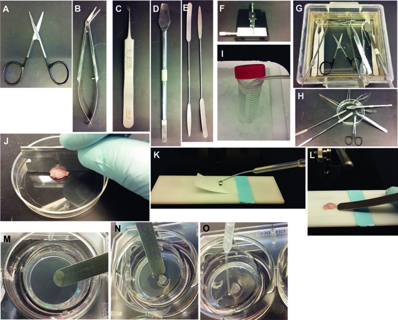
Fig. 1.
Setup for making organotypic slice cultures from P8 mouse brain. (a–e) Tools. (f) Tissue chopper. (g, h) Treatment of tools with 70% ethanol. (i) Bubbling dissection buffer with 90% O2/5% CO2 gas mixture and chilling it on ice. (j) Cutting the dissected brain into two hemispheres. (k) Preparation of the stage for slicing using Whatman paper to hold the brain in place. (l) Picking up sliced hemisphere with a spatula. (m, n) Sliding a slice onto the membrane of a Millicell insert. O: Removing extra dissection buffer from around the slice using a transfer pipet

