
Figure 2.
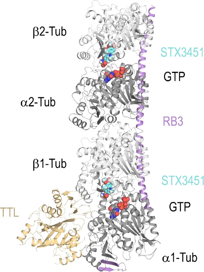
Overall view of the T2R-TTL-STX3451 complex structure. The α- and β-tubulin chains are in dark and light grey, respectively, TTL is in yellow/orange, and RB3 is in violet ribbon representation. The tubulin-bound STX3451 and the nonexchangeable guanosine 5′-triphosphate (GTP) molecules bound to α-tubulin are shown as sphere representations. The carbon atoms of STX3451 are colored in cyan.