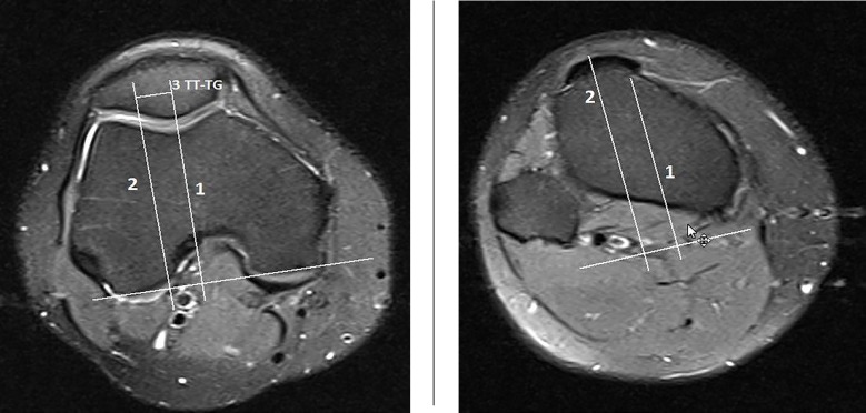
Figure 1.
axial image of knee joint at distal femur.note to posterior line was drawn at posterior border of condyles of femur. there is a vertical line at trochlear groove and the other parallel line along the tibial tuberosity that transfered to this level and distance between two lines indicate TT-TG

