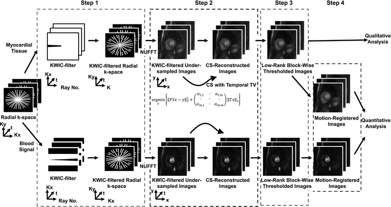
Figure 2:
A schematic of the image reconstruction pipeline involving 4 steps. In step 1, a KWIC filter was applied to the radial k-space. For the AIF image reconstruction, the k-space center was retained for the first ray and omitted for all the other rays. For the myocardial wall image reconstruction, the k-space center was retained for the last fifteen rays. In step 2, the undersampled images were reconstructed using CS with temporal total variation (TTV) as the sparsifying transform (30 iterations). A different regularization weight was used for the foreground and background. In step 3, three iterations of low-rank block-wise thresholding was performed to suppress the residual artifacts and noise. In step 4, in order to generate MBF maps, we performed motion correction using ANTS. For dynamic display of results after each of 3 steps (TTV, TTV + low-rank block-wise, TTV + low-rank block-wise + motion correction) in the reconstruction pipeline, see Supporting Information Video S1.

