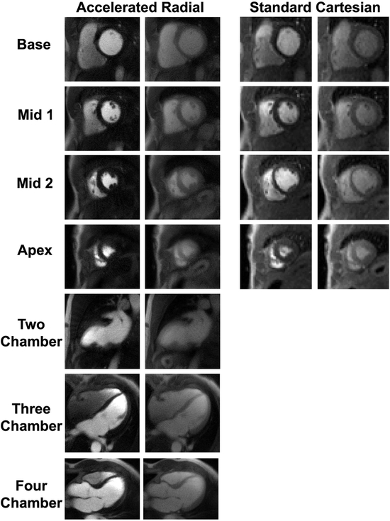
Figure 4:
Representative cardiac perfusion images obtained using the accelerated radial sequence (columns 1–2) and the standard perfusion sequence (columns 3–4) at peak enhancement in the blood pool (columns 1 and 3) and myocardial wall (columns 2 and 4). Using the proposed sequence, there was time left in the cardiac cycle to acquire three additional long-axis images. For dynamic display, see Supporting Information Video S2 for standard images and Supporting Information Video S3 for accelerated images.

