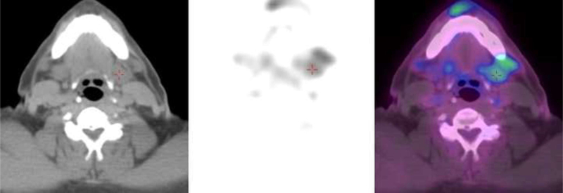
Figure 1:

Figure 1a: 18F-FDG PET/CT shows submandibular gland labeled by red cross. Suspicious lymph node is anterolateral to the red cross. Images represent a false positive detection of a preoperative metastatic cervical lymph node by 18F-FDG PET/CT. Although abnormal focal tracer uptake is found on imaging, no evidence of metastatic disease was found on histopathology. Figure 1b: CECT shows the same patient with normal anatomy, a large lymph node with fatty hilum, read correctly as negative for metastatic disease.
