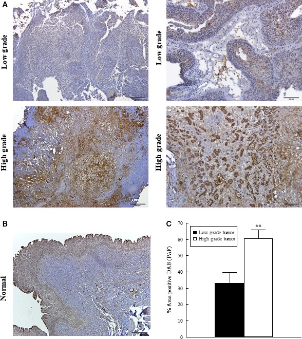
Figure 5.

Immunohistochemical expression of PAF in low‐ and high‐grade bladder cancer. Immunohistochemistry for PAF in low‐grade (top panels A, representative images) and high‐grade (lower panels A, representative images) tumors from smoker patients. PAF expression in normal human urothelial tissue (representative image, B). Quantification of PAF signal in tumor tissues (C). Values shown are means + SEM for three separate patient samples. **P < 0.01 when compared to controls. Scale bars: 200 μm.
